Signalment & History
Signalment:
- 8 weeks old
- Intact male
- Border collie
History:
- Just adopted
Presenting Complaint
- Here for initial health screening, deworming, and first puppy shot
- Left eye seems to be sunken in compared to the right
What is on your problem list?
- Microphthalmia
Physical Exam
- Temp: 101.5 F; Pulse: 70 bpm; Respiration: 25/min; MM: pink/moist CRT: 2 sec; BCS: 5/9; Eyes: microphthalmia OS; Ears: wnl; Mouth: wnl; LN’s: wnl; Cardio: wnl; Resp: wnl; Abd: wnl; GI: wnl; MS: wnl; CNS: wnl; Weight: 8 lbs (4 kg)
What diagnostics do you perform?
- To test for CEA, perform fundoscopy OS
- Scleral coloboma diagnosed
What is your assessment of this finding?
- This dog will likely lose vision in the left eye. Referral to ophthalmologist. Genetic testing recommended between visits.
What is your plan for this dog? How will you follow up with this patient?
- This dog should be monitored by an ophthalmologist. Ongoing communication with GP recommended.
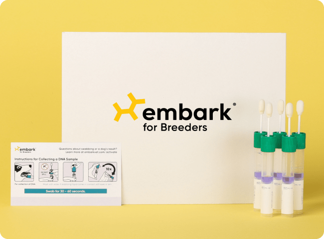
- Discussed ways to help dog adapt to decreased vision such as making sure the household is quiet, with normal daily routine, free from extraneous factors so that dog can familiarize self with house slowly. Guided leash walks recommended. Using this dog’s Embark results (revealing his at-risk result for CEA), the ophthalmologist could explain the ophthalmic defect.
Outcome
- While finding out this genetic result after ocular complications had already occurred did not change this dog’s outcome, the owners expressed great appreciation for the better understanding they had of their dog and his health. They now had an answer for why he lost vision in his left eye and could decide on a management plan with the ophthalmologist for his right eye.
Learn More: Diagnosing CEA
- CEA is present from birth, however, by three months of age the puppy fundus changes from its blue tapetal color to the adult green-yellow appearance. At the same time minor chorioretinal changes can be masked by the development of more pigment in the RPE, in which case affected animals are classified as so-called “go normal“ animals.
References/Additional Resources
- https://vcahospitals.com/know-your-pet/collie-eye-anomaly#:~:text=CEA%20may%20be%20diagnosed%20by,for%20a%20complete%20eye%20examination
- https://www.pethealthnetwork.com/dog-health/dog-diseases-conditions-a-z/collie-eye-anomaly-cea
- http://www.eyecareforanimals.com/conditions/inherited-eye-diseases-in-collies/
- https://youtu.be/rs1E54D6k14
* Embark is not necessarily affiliated with any of these websites or references, and does not necessarily endorse their content.


